
Sitemap
A list of all the posts and pages found on the site. For you robots out there, there is an XML version available for digesting as well.
Pages
Posts
Blog Post number 1
Published:
This is a sample blog post. Lorem ipsum I can’t remember the rest of lorem ipsum and don’t have an internet connection right now. Testing testing testing this blog post. Blog posts are cool.
publications
Orientation-Tuned Surround Suppression in Mouse Visual Cortex
Published in Journal of Neuroscience, 2014
- The firing rates of neurons in primary visual cortex (V1) are suppressed by large stimuli, an effect known as surround suppression. In cats and monkeys, the strength of suppression is sensitive to orientation; responses to regions containing uniform orientations are more suppressed than those containing orientation contrast. This effect is thought to be important for scene segmentation, but the underlying neural mechanisms are poorly understood. We asked whether it is possible to study these mechanisms in the visual cortex of mice, because of recent advances in technology for studying the cortical circuitry in mice. It is unknown whether neurons in mouse V1 are sensitive to orientation contrast. We measured the orientation selectivity of surround suppression in the different layers of mouse V1. We found strong surround suppression in layer 4 and the superficial layers, part of which was orientation tuned: iso-oriented surrounds caused more suppression than cross-oriented surrounds. Surround suppression was delayed relative to the visual response and orientation-tuned suppression was delayed further, suggesting two separate suppressive mechanisms. Previous studies proposed that surround suppression depends on the activity of inhibitory somatostatin-positive interneurons in the superficial layers. To test the involvement of the superficial layers we topically applied lidocaine. Silencing of the superficial layers did not prevent orientation-tuned suppression in layer 4. These results show that neurons in mouse V1, which lacks orientation columns, show orientation-dependent surround suppression in layer 4 and the superficial layers and that surround suppression in layer 4 does not require contributions from neurons in the superficial layers.
Recommended citation: Matthew W Self, Jeannette AM Lorteije, Joris Vangeneugden, Enny H van Beest, Mihaela E Grigore, Christiaan N Levelt, J Alexander Heimel, Pieter R Roelfsema. (2014). "Orientation-Tuned Surround Suppression in Mouse Visual Cortex." Journal of Neuroscience.
Download Paper
Activity in lateral visual areas contributes to surround suppression in awake mouse V1
Published in Current Biology, 2019
- Neuronal response to sensory stimuli depends on the context. The response in primary visual cortex (V1), for instance, is reduced when a stimulus is surrounded by a similar stimulus [1, 2, 3]. The source of this surround suppression is partially known. In mouse, local horizontal integration by somatostatin-expressing interneurons contributes to surround suppression [4]. In primates, however, surround suppression arises too quickly to come from local horizontal integration alone, and myelinated axons from higher visual areas, where cells have larger receptive fields, are thought to provide additional surround suppression [5, 6]. Silencing higher visual areas indeed decreased surround suppression in the awake primate by increasing responses to large stimuli [7, 8], although not under anesthesia [9, 10]. In smaller mammals, like mice, fast surround suppression could be possible without feedback. Recent studies revealed a small reduction in V1 responses when silencing higher areas [11, 12] but have not investigated surround suppression. To determine whether higher visual areas contribute to V1 surround suppression, even when this is not necessary for fast processing, we inhibited the areas lateral to V1, particularly the lateromedial area (LM), a possible homolog of primate V2 [13], while recording in V1 of awake and anesthetized mice. We found that part of the surround suppression depends on activity from lateral visual areas in the awake, but not anesthetized, mouse. Inhibiting the lateral visual areas specifically increased responses in V1 to large stimuli. We present a model explaining how excitatory feedback to V1 can have these suppressive effects for large stimuli.
Recommended citation: Joris Vangeneugden, Enny H van Beest, Michael X Cohen, Jeannette AM Lorteije, Sreedeep Mukherjee, Lisa Kirchberger, Jorrit S Montijn, Premnath Thamizharasu, Daniela Camillo, Christiaan N Levelt, Pieter R Roelfsema, Matthew W Self, J Alexander Heimel (2019). "Activity in lateral visual areas contributes to surround suppression in awake mouse V1." Current Biology.
Download Paper
Mouse visual cortex contains a region of enhanced spatial resolution
Published in Nature Communications, 2021
- The representation of space in mouse visual cortex was thought to be relatively uniform. Here we reveal, using population receptive-field (pRF) mapping techniques, that mouse visual cortex contains a region in which pRFs are considerably smaller. This region, the “focea,” represents a location in space in front of, and slightly above, the mouse. Using two-photon imaging we show that the smaller pRFs are due to lower scatter of receptive-fields at the focea and an over-representation of binocular regions of space. We show that receptive-fields of single-neurons in areas LM and AL are smaller at the focea and that mice have improved visual resolution in this region of space. Furthermore, freely moving mice make compensatory eye-movements to hold this region in front of them. Our results indicate that mice have spatial biases in their visual processing, a finding that has important implications for the use of the mouse model of vision.
Recommended citation: Enny H van Beest, Sreedeep Mukherjee, Lisa Kirchberger, Ulf H Schnabel, Chris van der Togt, Rob RM Teeuwen, Areg Barsegyan, Arne F Meyer, Jasper Poort, Pieter R Roelfsema, Matthew W Self (2021). "Mouse visual cortex contains a region of enhanced spatial resolution." Nature communications.
Download Paper
The essential role of recurrent processing for figure-ground perception in mice
Published in Science advances, 2021
- The segregation of figures from the background is an important step in visual perception. In primary visual cortex, figures evoke stronger activity than backgrounds during a delayed phase of the neuronal responses, but it is unknown how this figure-ground modulation (FGM) arises and whether it is necessary for perception. Here, we show, using optogenetic silencing in mice, that the delayed V1 response phase is necessary for figure-ground segregation. Neurons in higher visual areas also exhibit FGM and optogenetic silencing of higher areas reduced FGM in V1. In V1, figures elicited higher activity of vasoactive intestinal peptide–expressing (VIP) interneurons than the background, whereas figures suppressed somatostatin-positive interneurons, resulting in an increased activation of pyramidal cells. Optogenetic silencing of VIP neurons reduced FGM in V1, indicating that disinhibitory circuits contribute to FGM. Our results provide insight into how lower and higher areas of the visual cortex interact to shape visual perception.
Recommended citation: Lisa Kirchberger, Sreedeep Mukherjee, Ulf H Schnabel, Enny H van Beest, Areg Barsegyan, Christiaan N Levelt, J Alexander Heimel, Jeannette AM Lorteije, Chris van der Togt, Matthew W Self, Pieter R Roelfsema (2021). "The essential role of recurrent processing for figure-ground perception in mice." Science advances.
Download Paper
Unraveling the mechanisms of deep-brain stimulation of the internal capsule in a mouse model
Published in Nature Communications, 2023
- Deep-brain stimulation (DBS) is an effective treatment for patients suffering from otherwise therapy-resistant psychiatric disorders, including obsessive-compulsive disorder. Modulation of cortico-striatal circuits has been suggested as a mechanism of action. To gain mechanistic insight, we monitored neuronal activity in cortico-striatal regions in a mouse model for compulsive behavior, while systematically varying clinically relevant parameters of internal-capsule DBS. DBS showed dose-dependent effects on both brain and behavior: An increasing, yet balanced, number of excited and inhibited neurons was recruited, scattered throughout cortico-striatal regions, while excessive grooming decreased. Such neuronal recruitment did not alter basic brain function such as resting-state activity, and only occurred in awake animals, indicating a dependency on network activity. In addition to these widespread effects, we observed specific involvement of the medial orbitofrontal cortex in therapeutic outcomes, which was corroborated by optogenetic stimulation. Together, our findings provide mechanistic insight into how DBS exerts its therapeutic effects on compulsive behaviors.
Recommended citation: Bastijn JG van den Boom, Alfredo Elhazaz-Fernandez, Peter A Rasmussen, Enny H van Beest, Aishwarya Parthasarathy, Damiaan Denys, Ingo Willuhn (2023). "Unraveling the mechanisms of deep-brain stimulation of the internal capsule in a mouse model." Nature communications.
Download Paper
The direct and indirect pathways of the basal ganglia antagonistically influence cortical activity and perceptual decisions
Published in iScience, 2024
- The striatum, the main input nucleus of the basal ganglia, receives topographically organized input from the cortex and gives rise to the direct and indirect output pathways, which have antagonistic effects on basal ganglia output directed to the cortex. We optogenetically stimulated the direct and indirect pathways in a visual and a working memory task in mice that responded by licking. Unilateral direct pathway stimulation increased the probability of lick responses toward the contralateral, non-stimulated side and increased cortical activity globally. In contrast, indirect pathway stimulation increased the probability of responses toward the stimulated side and decreased activity in the stimulated hemisphere. Moreover, direct pathway stimulation enhanced the neural representation of a contralateral visual stimulus during the delay of the working memory task, whereas indirect pathway stimulation had the opposite effect. Our results demonstrate how these two pathways influence perceptual decisions and working memory and modify activity in the dorsal cortex.
Recommended citation: Enny H van Beest, Mohammed AO Abdelwahab, J Leonie Cazemier, Chrysiida Baltira, M Cassandra Maes, Brandon D Peri, Matthew W Self, Ingo Willuhn, Pieter R Roelfsema (2024). "The direct and indirect pathways of the basal ganglia antagonistically influence cortical activity and perceptual decisions." iScience.
Download Paper
Tracking neurons across days with high-density probes
Published in Nature Methods, 2024
- Neural activity spans multiple time scales, from milliseconds to months. Its evolution can be recorded with chronic high-density arrays such as Neuropixels probes, which can measure each spike at tens of sites and record hundreds of neurons. These probes produce vast amounts of data that require different approaches for tracking neurons across recordings. Here, to meet this need, we developed UnitMatch, a pipeline that operates after spike sorting, based only on each unit’s average spike waveform. We tested UnitMatch in Neuropixels recordings from the mouse brain, where it tracked neurons across weeks. Across the brain, neurons had distinctive inter-spike interval distributions. Their correlations with other neurons remained stable over weeks. In the visual cortex, the neurons’ selectivity for visual stimuli remained similarly stable. In the striatum, however, neuronal responses changed across days during learning of a task. UnitMatch is thus a promising tool to reveal both invariance and plasticity in neural activity across days.
Recommended citation: Enny H van Beest, Célian Bimbard, Julie MJ Fabre, Sam W Dodgson, Flóra Takács, Philip Coen, Anna Lebedeva, Kenneth D Harris, Matteo Carandini (2025). "Tracking neurons across days with high-density probes." Nature Methods.
Download Paper
An adaptable, reusable, and light implant for chronic Neuropixels probes
Published in eLife, 2025
- Electrophysiology has proven invaluable to record neural activity, and the development of Neuropixels probes dramatically increased the number of recorded neurons. These probes are often implanted acutely, but acute recordings cannot be performed in freely moving animals and the recorded neurons cannot be tracked across days. To study key behaviors such as navigation, learning, and memory formation, the probes must be implanted chronically. An ideal chronic implant should (1) allow stable recordings of neurons for weeks;(2) allow reuse of the probes after explantation;(3) be light enough for use in mice. Here, we present the ‘Apollo Implant’, an open-source and editable device that meets these criteria and accommodates up to two Neuropixels 1.0 or 2.0 probes. The implant comprises a ‘payload’module which is attached to the probe and is recoverable, and a ‘docking’module which is cemented to the skull. The design is adjustable, making it easy to change the distance between probes, the angle of insertion, and the depth of insertion. We tested the implant across eight labs in head-fixed mice, freely moving mice, and freely moving rats. The number of neurons recorded across days was stable, even after repeated implantations of the same probe. The Apollo implant provides an inexpensive, lightweight, and flexible solution for reusable chronic Neuropixels recordings.
Recommended citation: Célian Bimbard, Flóra Takács, Joana A Catarino, Julie MJ Fabre, Sukriti Gupta, Stephen C Lenzi, Maxwell D Melin, Nathanael O'Neill, Ivana Orsolic, Magdalena Robacha, James S Street, José M Gomes Teixeira, Simon Townsend, Enny H van Beest, Arthur M Zhang, Anne K Churchland, Chunyu A Duan, Kenneth D Harris, Dimitri Michael Kullmann, Gabriele Lignani, Zachary F Mainen, Troy W Margrie, Nathalie L Rochefort, Andrew Wikenheiser, Matteo Carandini, Philip Coen (2025). "An adaptable, reusable, and light implant for chronic Neuropixels probes." eLife.
Download Paper
DeepUnitMatch: tracking neurons across days in electrophysiology using Deep Neural Networks
Published in bioRxiv, 2026
- To understand neural processes such as learning or memory, we need to track the activity of populations of neurons at the level of single spikes and across days. Here, we leverage deep neural networks to build DeepUnitMatch, a software that reliably tracks individual neurons in high-density electrophysiological recordings across weeks. DeepUnitMatch uses only the spike waveforms of the neurons, and not their spiking patterns, and outperforms current solutions.
Recommended citation: Suyash Agarwal, Wentao Qiu, Kenneth D Harris, Enny H van Beest, Célian Bimbard (2026). "DeepUnitMatch: tracking neurons across days in electrophysiology using Deep Neural Networks." bioRxiv.
Download Paper
software
UnitMatch
Toolbox to track single units (electrophysiology) across recordings 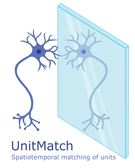
Bombcell
Automated curation and cell classification of spike-sorted electrophysiology data (Julie Fabre) 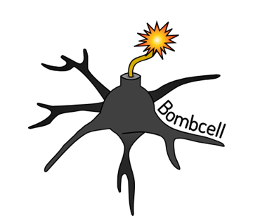
talks
teaching
Private tutor
private tutor, StudentsPlus, 2010
Private Tutor (via StudentsPlus Amsterdam) for high school students. Topics: Mathematics, Biology, Physics and Chemistry.
Statistics in R
Student assistent, University of Amsterdam, 2013
Student assistant for Bachelor of Psychobiology, University of Amsterdam. Topic: Statistics in R
UCL Neuropixels Course 2023
Online specialistic course, University College London, 2023
Neuropixels probes are transforming neurophysiology, and are now adopted by hundreds of laboratories worldwide. This free online course trained scientists to use these probes in their experiments, and to process their output. The course is supported by the Wellcome Trust. All course materials are freely available online.
UCL Neuropixels Course 2024
Online specialistic course, University College London, 2024
UCL Neuropixels Course 2025
Online specialistic course, University College London, 2025
Open Research
Course developer / lecturer, University College London, 2025
Four-week online workshop series designed to introduce PhD students and newcomers across the faculty to the principles and practices of open research. I organized session 3: Open Access Publishing and Open Data
Lecture - Organisation of the visual system
Lecturer, University College London, 2026
Lecture + additional tutorial on ‘Organisation of the ascending and descending neural pathways of the visual system I’ for the module ‘Introduction to Human Sensory systems’ at the Institute of Ophthalmology - UCL.
Lecture - Vision & Navigation
Lecturer, University College London, 2026
Lecture on ‘Vision and Navigation’ for the module ‘Behavioural and Systems Neuroscience’ in the master Neuroscience - UCL.